Yordanova, J., Kolev, V. (1996). Developmental changes in the alpha response system. Electroencephalography and clinical Neurophysiology, 99: 527-538. Copyright © 1996 Elsevier Science Ireland Ltd. 

   
Developmental Changes in the Alpha Response System
 

Juliana Y. Yordanova a,b,*, Vasil N. Kolev a,b
aBrain Research Institute, Bulgarian Academy of Sciences, 1113 Sofia, Bulgaria
bInstitute of Physiology, Medical University of Lübeck, D-23538 Lübeck, Germany
 
Accepted for publication:  31 May 1996
 
    *Corresponding author: Institute of Physiology, Bulgarian Academy of Sciences,
      Acad. G. Bonchev str. bl. 23, 1113 Sofia, Bulgaria. Tel.: (359) 2-713-37-49 Fax: (359) 2-738-469
      email: jyord@iph.bio.acad.bg


Abstract
 
    Evoked and event-related brain potentials (ERPs) may be regarded as originating from the reorganization of the spontaneous EEG rhythms. The main objective of the present research was to study the alpha responses in 6-11 year-old children to determine whether the ability to reorganize alpha activity after external stimulation demonstrates developmental changes that could reflect variations in information processing with increased age.  A total of 50 children aged 6-11 years, divided into 5 age groups, and 10 young adults were assessed in a passive and an oddball condition. Alpha responses in the passive and non-target ERPs at Fz, Cz and Pz were analyzed to assess quantitatively the repeatability (phase-locking) of the evoked alpha oscillations. The alpha responses in 6-11 year-old children were different from those in adults:  (1) Adults had significantly lower amplitude and stronger phase-locking than children;  (2) Adults had maximal alpha amplitudes and phase-locking over the vertex, whereas children displayed maximal responses over the parietal site;  (3) The phase-locking of eldest (10-11 year-old) children was as strong as in adults. These findings indicate that the alpha response system is functionally involved in 6-11 year-old children, though its development is not complete at the age of 11, and the magnitude and the phase-locking parameters may relate to different functional aspects of the alpha response system. Thus, younger children do produce alpha responses during information processing, but are not able to engage this system as strongly as older children and adults. Copyright © 1996 Elsevier Science Ireland Ltd.
 
Keywords:   Alpha response; Phase-locking; Event-related potentials; Auditory evoked potentials; Children; EEG


 1. Introduction
 
    After external and/or internal stimulation, oscillatory potentials in different frequency bands can be recorded from the brain (Basar and Bullock 1992; Pantev et al. 1994). These oscillatory responses are called evoked or induced rhythmicities (Basar 1980, 1992; Galambos 1992; Bullock 1992; Bullock and Achimowicz 1994). Many investigators regard the evoked and event-related brain potentials (EPs, ERPs) as originating from the reorganization of the rhythmic activity in the spontaneous EEG, with specific time-domain components proposed to reflect externally or internally evoked synchronization, stabilization, selective enhancement, and phase-reordering of various frequencies in the ongoing EEG (Basar and Özesmi 1972; Davis 1973; Sayers et al. 1974; Davis 1976; Parvin et al. 1980; Basar 1980, 1992; Stampfer and Basar 1985).
 
    The oscillatory activity in the alpha range in the first 250-300 ms after stimulation defines the alpha response or the alpha frequency component of the evoked or event-related brain potential. The alpha response is assumed to be the most fundamental and almost invariant component of brain neuroelectric activity, since a number of cortical and subcortical structures manifest the ability to respond to stimuli in different modalities with damped oscillations in the alpha frequency band (for reviews see Basar 1980, 1992; Galambos 1992). Cellular recordings also have demonstrated alpha responses in subcortical and cortical neurons (e.g., Llinás 1988; Steriade et al. 1990; Dinse et al. 1990). It has been suggested that alpha networks with similar design are distributed in various brain structures where they comprise the alpha system of the brain and generate spontaneous alpha oscillations in addition to their ability to respond in the alpha range (Lopes da Silva et al. 1973; Lopes da Silva 1987; Basar 1980, 1992). With regard to the possible functional meaning of the alpha rhythm, recent results indicate strongly that alpha is not just a spontaneous background EEG rhythm but appears related to primary sensory processing (Basar and Schürmann 1994), motor behaviour (Pfurtscheller and Klimesch 1992), memory processes (Klimesch et al. 1993), and anticipation (Basar et al. 1989). Thus, the alpha frequency component of the event-related brain potentials  may reflect important aspects of information processing by the brain.
 
    Although developmental changes in the time domain EPs and ERPs have been intensively studied (e.g., Kurtzberg et al. 1984; Courchesne 1983, 1990; Friedman 1991; Polich et al. 1990), no information has been provided about the development of the frequency components of EPs and ERPs in children. The age-dependent variations in alpha responses of children have been described only recently. In contrast to adults, three-year-old children did not manifest consistent alpha frequency components in the evoked auditory and visual potentials (Basar-Eroglu et al. 1994; Kolev et al. 1994). Also, those children who did produce alpha responses, were not able to synchronize them with the moment of stimulus occurrence. These findings imply that the alpha response may undergo specific variations from childhood to adulthood, although the precise developmental course is not known. The present study was designed to assess how alpha response system functioning changes in the course of development as reflected by age differences in the alpha frequency components of ERPs. Another report discusses the theta response data from the same groups of children (Yordanova and Kolev, in press). Here, the following two hypotheses were evaluated.
 
    (1) The developmental changes in the spontaneous EEG in children have been previously described, with the consistent finding that the age-related reduction of the spontaneous EEG activity in the slower (delta, theta) frequency bands is accompanied by an increase in the faster (alpha and beta) frequency bands (Petersén and Eeg-Olofsson 1971; Matoušek and Petersén 1973; John et al. 1980; Matthis et al. 1980; Katada et al. 1981; Dubrovinskaya 1985; Gasser et al. 1988a; Gasser et al. 1988b; Niedermeyer 1987). The developmental dynamics of the alpha band power in the spontaneous EEG imply that the activity and/or the number of neuronal populations involved in the alpha system changes with age, such that the magnitude of the alpha responses would also vary as age increases. This hypothesis was assessed by examining the age-related dynamics of alpha response amplitude in single-sweep and averaged potentials.
 
    (2) The second hypothesis is that the capability of reorganizing the ongoing EEG and producing repeatable and stable alpha patterns during stimulus processing is related closely to brain development. To study the phase-locking phenomena, the congruence between consecutive single alpha responses was analyzed using  an original method that allows quantification of the number of phase-locked alpha waves present in the single-sweep responses (Kolev and Daskalova 1990).
 
    Alpha frequency components of the auditory event-related potentials were studied in 6-11 year-old children and in young adults. Amplitude and phase-locking of the alpha responses were examined with respect to their age- and topography dependence, with the age factor expected to produce variations in evoked alpha amplitudes and a concomitant change in phase-locking that can be observed distinctly with appropriate quantitative methods. To approach the question of how the maturation of the spontaneous alpha rhythm contributes to the evoked alpha response, the relationship between alpha-band power of the pre-stimulus EEG and single-sweep parameters also was studied.


2. Method
 
2.1. Subjects
 
    A total of 50 healthy children from 6 to 11 years of age served as subjects, with ten adults from 20 to 30 years of age also assessed. The ages of children ranged between 72 and 132 months, and were divided into 5 age groups consisting of 10 subjects each as indicated in Table 1. Children were obtained from local schools and adult subjects were volunteers, primarily students from the Medical University, Sofia. All subjects were right-handed, denied any history of neurologic, psychiatric disorders, or hearing problems. Interviews with teachers and parents of the children revealed no signs of attentional, behavioural disturbances, or learning problems. The children were of similar socio-economic status and had normal and above IQ scores. All subjects were paid for their participation in the experiment.
 
 
Table 1
 
Distribution of the subjects studied according to age
 
Children         Adults
6-7 years 7-8 years 8-9 years 9-10 years 10-11 years 20-30 years
Age (years) 6.50 7.60 8.50 9.30 10.60 24.10
SD (months) 4.65 3.25 4.68 3.03 4.43 44.40
 
 
2.2. Stimuli and procedure
 
    The subjects were assessed in a dimly lit, electrically shielded room and were monitored by means of a closed-loop TV and interphone system. Children were given enough time before the recording sessions to become acquainted with the environment.
 
    The auditory stimuli were generated by an IBM-AT386 computer, filtered, amplified, and reproduced by a loudspeaker in a free-sound field. All stimuli were presented at 60 dB SPL, with a duration of 50 ms (r/f 10 ms). The stimuli were delivered with random inter-stimulus intervals (3.5-6.5 s) in two different task situations: (1) Tone bursts of 800 Hz frequency (N = 50) were presented in a passive listening condition, with subjects instructed to relax silently keeping their eyes closed; (2) Frequent non-target stimuli were presented in an oddball task. During the oddball condition, 100 high and low frequency tones (1200 Hz and 800 Hz) were presented randomly, with P = 0.75 for the high tones, and P = 0.25 for the low tones. Subjects were required to press a button with their dominant hand as quickly and accurately as possible in response to the low tones. The rationale for studying two types of task-irrelevant alpha responses to passive and non-target stimuli is to ensure that any age-related differences were not due to the specific requirements in a single paradigm. The target oddball data will be reported elsewhere.
 
 
2.3. Data collection and processing
 
 
2.3.1. Electrodes
 
    The EEG data were recorded with Ag-AgCl disc electrodes placed on midline frontal, central and parietal sites (Fz, Cz, Pz), with linked-mastoids as the reference. The ground electrode was positioned on the forehead. The electrooculogram (EOG) was recorded bipolarly with electrodes placed below and at the outer canthus of the left eye. Electrode impedance did not exceed 10 KOhms.
 
 
2.3.2. EEG recording, ADC, and data storage
 
    EEG was amplified with band limits of 0.5 and 70 Hz by means of a Nihon Kohden Electroencephalograph (model EEG-4314F). Stop-band filtering (band limits 48 - 52 Hz) was used for eliminating interference. The amplified EEG analog signals were digitized with a sampling frequency of 250 Hz and stored on the computer hard disk, with an epoch length of 1024 ms pre- and 1024 ms post-stimulus.
 
 
2.3.3. Artifact rejection
 
    The stored raw single sweeps were analyzed visually off-line to eliminate EEG segments contaminated with blink, muscular or any other type of artifact activity. Also, any EEG or EOG trial exceeding ±50 µV was excluded from further analysis. Thus, the number of artifact-free sweeps analyzed for each subject in each condition was between 40 and 50.
 
 
2.4. Data Analysis
 
    ERP component analysis in both the time and frequency domains was employed, which included selective averaging, calculation of the amplitude-frequency characteristics, and response adaptive digital filtering. In addition, an analysis of single alpha responses was performed. Fig. 1 presents a schematic illustration of  the methods used and their relationship to each other. A brief description of the analysis procedures follows.
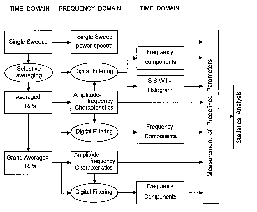
Fig. 1. Schematic presentation of the methods and procedures used to perform time and frequency domain analysis at levels of single sweeps, averaged and grand averaged ERPs, and their relationship.
 
 
2.4.1. Amplitude frequency-characteristics (AFCs) computation
 
    After the selective averaging auditory ERPs were transformed to the frequency domain with fast Fourier transform (FFT) to obtain the amplitude-frequency characteristics :

where c(t) is a step function of the system (in this case, the evoked or event-related potential), and , where f  is the frequency of the input signal. Although this transform is valid only for linear systems, it can be applied  to nonlinear systems as a first approach, since errors from system nonlinearities are smaller than errors resulting from the length of measurements and rapid transitions in brain activity. For details on the method of transient response frequency characteristics see Basar (1980) and Röschke et al. (1995).
 
 
2.4.2. Digital filtering
 
    Response adaptive digital filtering (Basar and Ungan 1973; Basar 1980), which provides zero phase shift, was used to compute the ERP frequency components. Individual single sweeps in each series as well as the averaged ERPs were digitally pass-band filtered in the frequency range of 8-15 Hz to cover individual variations in alpha peaks of the AFCs of both children and adults.
 
 
2.4.3. Single sweep analysis
 
    Amplitudes and phase-locking of single alpha responses were analyzed separately to examine their possible independent changes with increasing age. For a quantitative evaluation of the phase-locking for single sweeps that were filtered in the alpha range, a modification of the Single Sweep Wave Identification (SSWI) method was applied (Kolev and Daskalova 1990). The analysis procedure included the steps described below, which are illustrated schematically in Fig. 2.
 
    First, extrema (minima and maxima) were identified in the filtered (8-15 Hz) single sweeps that were chosen for selective averaging in each of the experimental series. Amplitude and latency values of the identified extrema were stored. This step is illustrated in Fig. 2a where three representative single sweeps are shown (left panel), with the detected points along the time axis, presented without the signals (right panel).
 
    Second, a histogram of the number of phase-locked single alpha waves that occur in the analysis epoch (single sweep wave identification histogram, SSWI-histogram) was constructed. To perform data reduction, the analysis epoch was divided into time intervals of 8 ms. For each time interval, the sum of the number of the identified extrema was calculated, the maxima taken with plus sign and the minima taken with minus sign. Hence, the number of the phase-locked waves in the consecutive single sweeps for each 8 ms interval was determined, and the obtained value was assigned to the corresponding histogram bar (right bottom panel of Fig. 2a). The histogram thus reflects the stability in phase of the single sweep waves time-locked to stimulus independent of their amplitudes. A typical SSWI-histogram of an adult subject is shown in Fig. 2b. The strong phase-locking of the alpha responses in the first 250-300 ms after stimulus presentation is clear.
 
    Third, quantitative evaluation of single sweep phase-locking was performed. The SSWI-histogram was normalized by dividing the values by the number of single sweeps included. The sum of absolute bar values (integral) of the normalized SSWI-histogram was calculated for the time window 0-300 ms post-stimulus, thus quantifying the  information about the strength of single sweep phase-locking.
 
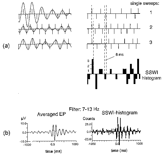
Fig. 2. SSWI method. (a) Three single sweeps (1, 2, 3) filtered in the alpha range (left), the identified extrema presented by their locations with bars equal to ±1 (right), and the sums of bars in time intervals of 8 ms building the SSWI-histogram. (b) A typical result from an adult subject:  averaged evoked potential filtered in the alpha range (left) and the corresponding SSWI-histogram (right). Number of single sweeps averaged and used for SSWI-histogram building is 96. Stimulus is presented at 0 ms.
 
 
2.4.4. Power spectral density of the pre-stimulus EEG
 
    The power spectral density functions were calculated for each artefact-free pre-stimulus epoch (-1000, 0 ms) using the fast Fourier transform algorithm and then averaged.
 
 
2.5. Statistical Analysis
 
    For the purposes of statistical analysis the following parameters of the alpha frequency component in ERPs and the pre-stimulus EEG were measured in the filtered (8-15 Hz) averaged and single sweep potentials for each lead and condition: (1) maximal peak-to-peak amplitude in the time window from 0 to 300 ms after stimulus in averaged filtered ERPs; (2) mean value of the maximal peak-to-peak amplitudes of all artifact-free single-sweep ERPs in the same time window (0 - 300 ms); (3) sum of absolute bar values (integral) of the normalized SSWI-histogram within the time window 0 - 300 ms post-stimulus; (4) absolute band power (X) in the range of 8-15 Hz of the pre-stimulus EEG, log-transformed according to the formula Y = log10(X) (Gasser et al. 1982, 1988a). Individual measurements of each parameter in each condition were subjected to a repeated-measures analysis of variance with one between-subjects variable, Age (6 levels, corresponding to each age group, 6-7, 7-8, 8-9, 9-10, 10-11, Adults), and one within-subjects variable, Electrode Location (Fz, Cz, Pz). The Greenhouse-Geisser correction procedure was applied to factors with repeated-measures. The original df and the probability values from the reduced df are reported here. Also, results from testing single effects and contrasts not presented in tables, are regarded as significant only if the probability values were smaller than 0.05. A similar analysis was carried out for the log- transformed absolute alpha-band power values of the pre-stimulus EEG recorded in the passive condition. To verify the significance of Age x Electrode interactions found for amplitude measures, analyses were redone after row data were MinMax normalized (McCarthy and Wood, 1985).


3. Results
 
 
3.1. Time domain ERPs
 
    Fig. 3 illustrates that: (1) The ERPs of children and adults were characterized by N1, P2, N2, and P3 components, with a stable configuration of the components in the waveforms achieved after the age of 6-7 years.  (2) At Pz, the component structure of ERPs in children was similar to that observed for adults, whereas at Fz and Cz a late negative wave N400-700 developed in children that was not expressed in adults. A detailed analysis of the time domain components is presented elsewhere (Yordanova et al. 1992).
 
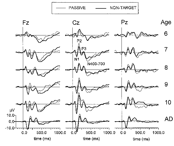
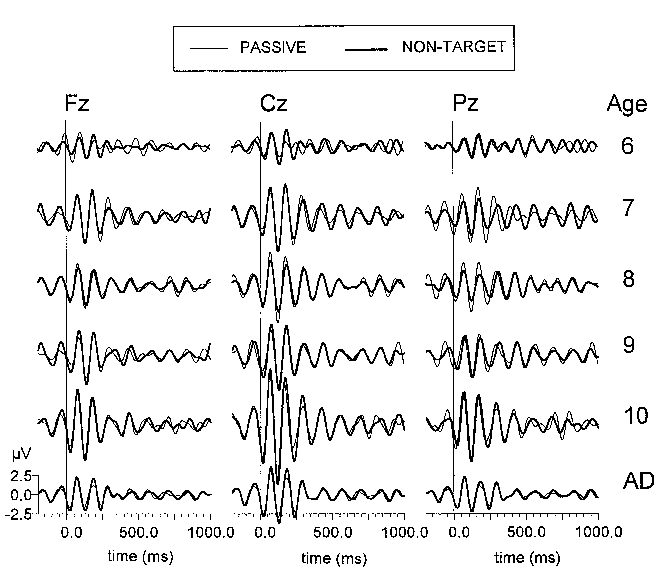
Fig. 3. Grand average passive (thin line) and non-target ERPs (thick line) at three electrode locations (Fz, Cz, Pz) of six age groups (6: 6-7 year-olds, 7: 7-8 year-olds, 8: 8-9 year-olds, 9: 9-10 year-olds, 10: 10-11 year-olds, AD: adults). Each age group consists of 10 subjects. Stimulus is presented at 0 ms.
 
 
3.2. Amplitude-frequency characteristics of ERPs
 
    Fig. 4 shows the AFCs of six representative subjects from each age group and illustrates that (1) peaks in the alpha range were present in both children and adults, and (2) the AFCs of children were different from those of adults with respect to the number of identifiable peaks. The AFCs of adults were typically characterized by a broad compound response covering the range of the theta and alpha frequencies (4-12 Hz) and peaking at 6-9 Hz. In younger children, distinct peaks were detected in the delta, theta, and alpha ranges. The number of separable peaks in the AFCs decreased with increasing age in children.
 
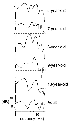
Fig. 4. Amplitude-frequency characteristics for 6 representative subjects at 6, 7, 8, 9, and 10 years of age and an adult, calculated from the passive ERPs recorded at Cz. Along the y-axis:  20log|| (dB).
 
 
3.3. Alpha responses in the ERPs
 
    Results from the statistical analysis are presented in Table 2, with univariate F-contrasts of differences between the age groups shown in Table 3. Fig. 5 illustrates alpha responses in grand average ERPs, while Fig. 6 displays representative individual data to visualize the specific information reflected by each of the three analyzed parameters. The main age effects are presented in Fig. 7, with the mean group values shown in Fig. 8.
 
Fig. 5. Grand average passive (thin line) and non-target (thick line) ERPs at three electrode locations (Fz, Cz, Pz) filtered in the alpha (8-15 Hz) range. The different age groups are designated in the same manner as in Fig. 3. Each age group consists of 10 subjects. Stimulus is presented at 0 ms.
 
 
Table 2
 
Summary of results from repeated measures analyses of variance of alpha response measurements for the passive and non-target ERPs
 
Parameters Source (df) Passive   Non-target  
F P F P
Aaver Age (5,54)   3.5   0.01   4.1 <0.001
Electrode (2,108)   9.2 <0.001 24.7 <0.001
A x E (10,108)   1.8   0.06   3.1 <0.001
Asin Age (5,54)   5.3 <0.001   4.9 <0.001
Electrode (2,108) 22.7 <0.001 26.1 <0.001
A x E (10,108)   2.3   0.04   2.2   0.05
Np Age (5,54)   3.4   0.01   6.1 <0.001
Electrode (2,108) 24.9 <0.001 47.4 <0.001
A x E (10,108)   4.3 <0.001   1.7   0.09
 
Aaver, amplitude of averaged alpha response; Asin, amplitude of single sweep alpha response; Np, normalized number of phase-locked alpha waves. Variables:  Age (6-7, 7-8, 8-9, 9-10, 10-11 year old children; adults) and Electrode location (Fz, Cz, Pz).
 
 
Table 3
 
Summary of univariate F-contrasts
 
Contrasts Passive           Non-target          
(df  1,54) Aaver Asin Np Aaver Asin Np
F P F P F P F P F P F P
6 vs 7
-
-
-
-
-
-
-
-
-
-
-
-
6 vs 8
-
-
-
-
-
-
-
-
-
-
-
-
6 vs 9
-
-
-
-
-
-
-
-
-
-
-
-
6 vs 10
9.5
<0.001
-
-
7.4
0.01
15.9
<0.001
-
-
10.9
<0.001
6 vs AD
-
-
15.6
<0.001
14.1
<0.001
-
-
10.7
<0.001
22.4
<0.001
7 vs 8
-
-
-
-
-
-
-
-
-
-
-
-
7 vs 9
-
-
-
-
-
-
-
-
-
-
-
-
7 vs 10
-
-
-
-
-
-
6.3
0.02
-
-
5.9
0.01
7 vs AD
5.2
0.003
13.8
<0.001
6.7
0.01
-
-
15.5
<0.001
15.4
<0.001
8 vs 9
-
-
-
-
-
-
-
-
-
-
-
-
8 vs 10
3.6
0.06
-
-
-
-
7.6
0.01
7.8
0.03
4.2
0.05
8 vs AD
3.9
0.05
9.9
<0.001
5.7
0.02
-
-
5.8
0.02
10.9
<0.001
9 vs 10
4.6
0.04
-
-
-
-
9.2
<0.001
-
-
4.4
0.04
9 vs AD
-
-
8.7
<0.001
5.3
0.03
-
-
8.4
0.01
12.6
<0.001
10vs AD
14.6
<0.001
21.4
<0.001
-
-
14.4
<0.001
20.4
<0.001
-
-
 
Age groups: 6, 6-7;  7, 7-8;  8, 8-9;  9, 9-10;  10, 10-11 year old children; AD, adults. Parameters are designated in the same manner as in Table 2. -, no significant difference, P>0.05.
 
Fig. 6. Averaged filtered ERPs (a), superimposed filtered single sweeps (b), and the corresponding SSWI-histograms (c) for six representative subjects at 6, 7, 8, 9, and 10 years of age, and an adult. Filter cut-off frequencies used are 8 and 15 Hz. All recordings are from the passive listening condition at Cz. Stimulus occurs at 0 ms.
 
 
3.3.1. Age and electrode effects:  alpha responses in average ERPs
 
    A significant main effect for Age was obtained for both conditions (Table 2), mainly due to the highest amplitudes in 10-11 year-old children (Table 3). As  illustrated in Fig. 5, Fig. 6a, and Fig. 7a, although an amplitude increase occurred after the age of 6-7 years, a second significant enhancement was found at the age of 10-11 years. Much lower alpha amplitudes were observed for adult subjects. Average alpha responses were highest over the vertex site (Fig. 8a). Further results from exploring Age x Electrode interaction will not be presented, since single-sweep analyses were in the focus of interest.
 
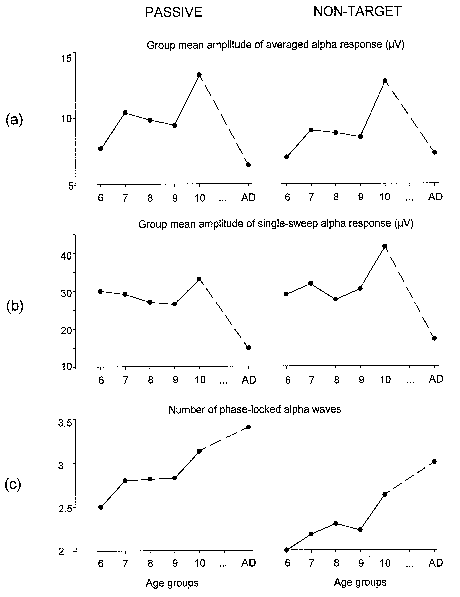
Fig. 7. Group mean amplitude of alpha responses in averaged passive and non-target ERPs (a), group mean amplitude of single sweep alpha responses (b), and group means of  the number of phase-locked alpha waves as calculated from normalized SSWI-histograms (c). All amplitude values are in µV, with age groups designated as in Fig. 3. Left column - passive, right column - non-target ERP.
 
 
3.3.2. Age and electrode effects:  single-sweep alpha amplitudes
 
    A significant main effect of the Age factor on the amplitudes of single-sweep alpha responses was found (Table 2), which is illustrated in Fig. 6b and Fig. 7b. No significant differences between groups of children were revealed (Table 3), with adult subjects displaying significantly lower amplitudes than each of the children groups. The single alpha responses were maximal at Pz because of the specific topography in children groups that was different in adults (Fig. 8b). The significance of the Age x Electrode interactions was confirmed for both the passive ERPs (F(10,108) = 2.45, P = 0.03) and non-target ERPs (F(10,108) = 2.3, P = 0.05) after the MinMax normalization. Testing single effects revealed that the electrode factor was significant in each group and condition (F(2,18) >= 3.15, P <= 0.05), except for the 8-9 year-olds in the passive condition. In adults, the single alpha responses were significantly higher at Cz than at Fz and Pz (univariate F-contrasts Cz vs. Fz/Pz for each of the passive or non-target ERPs, F(1,9) >= 6.71, P <= 0.05). In each group of children, the single alpha responses were maximal at Pz, with values at Fz significantly smaller than at Cz and Pz (Fz vs. Cz/Pz for each group in each condition, F(1,9) >= 6.17, P <= 0.05), and values at Cz significantly smaller than at Pz for 6-7 and 7-8 year olds (Cz vs. Pz in each condition, F(1,9) >= 5.9, P <= 0.05).
 
 
3.3.3. Age and electrode effects:  alpha response phase-locking
 
    A significant main effect of the Age factor on alpha response phase-locking was obtained for both conditions (Table 2), which is illustrated  in Fig. 6c and Fig. 7c. Although an increase in the phase-locking was evident after the age of 6-7 years, a significant increase was observed for the 10-11 year-old children that was followed by a further increase in adults (Table 3). No significant difference was found between the 10-11 year-olds and adults. For the non-target ERPs, 10-11 year-olds manifested alpha response phase-locking that was significantly higher than in the younger (6-9 year-old) children groups. Fig. 8c shows that single alpha responses were phase-locked primarily over the vertex site. In both conditions, the adults produced a significant Cz vs. Fz/Pz difference (Cz vs. Fz/Pz, F(1,9) >= 7.16, P <= 0.03). In contrast, no topographic differentiation was found in 6-7 year-old children in either condition. In the groups of 7-11 year-old children, a significant Cz vs. Pz difference was obtained (for each group and condition F(1,9) >= 9.9, P <= 0.01), and a trend for a strong frontal phase-locking was additionally revealed in 7-9 year-old children as indicated by the higher number of phase-locked waves at Fz than at Pz in these age groups (F(1,9) >= 9.4, P <= 0.05). However, the eldest children manifested an area-specific pattern of alpha wave phase-locking that was identical to that in adults.
 
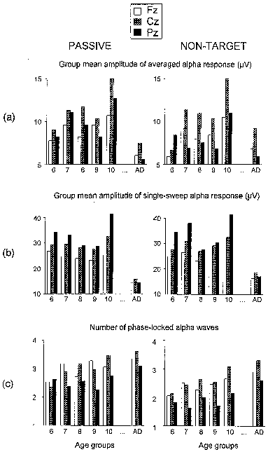
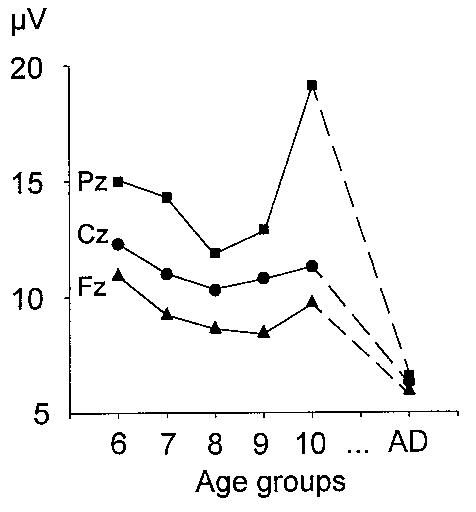
Fig. 8. Group mean amplitude of averaged alpha response (a), group mean amplitude of single sweep alpha response (b), and group means of the number of phase-locked alpha waves as calculated from normalized SSWI-histograms (c) versus age at three electrode locations (Fz, Cz, Pz). All amplitude values are in µV, with age groups designated as in Fig. 3. Left column - passive, right column - non-target ERP.
 
 
3.4. Absolute alpha band power of pre-stimulus activity
 
    Fig. 9 illustrates that the absolute band power of the pre-stimulus alpha activity was lowest in adults, with no differences obtained between groups of children (Age, F(5,54) = 8.3, P < 0.001). All age groups displayed maximal alpha power over the parietal area (Electrode, F(2,108) = 69.4, P < 0.001), with no significant interaction between age and electrode found after MinMax normalization (Age x Electrode, F(10,108) = 0.72, P > 0.1).
 
    Pearson correlation coefficients calculated for each lead were used to evaluate the relationship between pre-stimulus and evoked alpha activity. The correlation was positive and very strong for pre-stimulus alpha band power and single-sweep amplitude (Fz, r = 0.90; Cz, r = 0.86; Pz, r = 0.92; P < 0.001), but negative and much weaker for  pre-stimulus alpha power and phase-locking (Fz, r = –0.33; Cz, r = –0.39; Pz, r = –0.36; P < 0.02). No correlation was found between single alpha response amplitude and phase-locking.
 
Fig. 9. Mean group amplitude of the pre-stimulus alpha band activity in the passive condition at Fz, Cz and Pz. Age groups are designated as in Fig. 3.


4. Discussion
 
    In the present study, it was hypothesized that the amplitudes of average and single-sweep alpha responses to auditory stimuli  would differ between 6-11 year-old children and adults, and would undergo developmental changes with increasing age in children. Also, it was expected that the ability to reorganize alpha activity after external stimulation and to produce repeatable alpha patterns might be related closely to the processes of brain development with age. Major results demonstrated that: (1) Alpha responses were present in the ERPs of 6-11 year-old children. (2) Alpha responses in children differed significantly from those in adults who manifested lower single sweep amplitudes and stronger phase-locking. Furthermore, alpha responses  in adults were highest in amplitude and strongest in phase-locking over the mid-central brain area, whereas the maximal single-sweep alpha amplitudes in children were obtained over the parietal site. (3) Major developmental changes in alpha response system as reflected by single sweep parameters were revealed for the ability to synchronize with stimulus evoked alpha waves, since the phase-locking but not the amplitudes of single alpha responses increased significantly within groups of children. (4) Alpha response amplitude and phase-locking manifested specific and differential relationships with the power of the pre-stimulus alpha activity.
 
 
4.1. Alpha responses to auditory stimuli in children
 
    Enhanced alpha oscillations within 0-300 ms after auditory stimulation have been described previously in adults (Basar 1980; Særmark et al. 1992; Basar and Schürmann 1994). The present results from the adult subjects are in accordance with these observations and demonstrate additionally that the evoked alpha activity is mostly expressed at the vertex site. This topography pattern, especially of the phase-locking,  indicates that a specific organization of the alpha frequency component is present in relation with external auditory stimulation in adults.
 
    Alpha responses to auditory stimuli also were revealed in children aged from 6 to 11 years, because peaks in the alpha frequency range in the AFCs were found, and  enhanced and synchronized alpha oscillations were obtained after digital filtering of the ERPs in the 8-15 Hz frequency range (Fig. 6). In contrast, those three-year-old children in the studies of Basar-Eroglu et al. (1994) and Kolev et al. (1994) who had no developed spontaneous alpha activity, did not produce alpha responses to auditory and visual stimuli. Hence, the presence of developed pre-stimulus and evoked alpha activity in 6-11 year-old children gives a further evidence for the validity of the rule for brain response excitability: If a brain structure can generate intrinsic activity in a given frequency channel, then this structure also can respond to sensory stimulation in the same frequency channel (Sato et al. 1971, 1977; Basar 1980).
 
 
4.2. Alpha responses of children are different from adults
 
    The differences between the evoked alpha activity in children and adults suggest that the alpha response system in 6-11 year-old children might operate in a different mode than in adults, such that adults produce lower in amplitude but strongly synchronized responses. Single alpha response amplitudes and absolute alpha band power in the pre-stimulus EEG followed the same age-related changes and also manifested a strong relationship. This finding indicates that the maturation of the spontaneous alpha activity contributes to the age-related alterations in the magnitude of the alpha response. Physical factors like skull size may not be entirely responsible for the observed age-dependent differences in the alpha response amplitude because though the head circumference and head diameter have been reported to increase with age (Meredith 1971; Polich et al. 1990), (1) no significant amplitude differences were revealed among children groups examined here, and (2) the head size in children does not affect the time-domain ERP components consistently (Polich et al. 1990), nor the relationship between P3 and EEG (Intriligator and Polich 1995). Therefore, it may be concluded that due to a decrease in the spontaneous alpha power, adults generate alpha activity of lower amplitude than children do after auditory stimulation.
 
    More importantly, a shift in the maximal alpha activity from the parietal to the central site was present during auditory stimulus processing in adults. This ability to reorganize spatially and focus the maximal alpha power to the central site appears not yet developed even in children at 10-11 years of age, since they responded with maximal alpha amplitudes over these areas where the pre-stimulus alpha activity was mostly expressed.  Thus, the maturation of the 8-15 Hz response magnitude and scalp topography appears not complete at 11 years. However, the lack of difference between 10-11 year-olds and adults with regard to both the amount and topography of phase-locked alpha waves suggests that the mature level of alpha response phase-locking might be achieved at the age of 10-11 years. The possible implications of these findings are discussed below.
 
 
4.3. Developmental changes in the alpha response system
 
    The main developmental change in the alpha response system was found for the ability to produce repeatable and stable alpha wave packets after external (auditory) stimulation. The age-related increase in the amount of phase-locked alpha waves was discontinuous and also weakly related with the pre-stimulus alpha-band power. Although correlations with cognitive abilities of children were not made in the present study, it may be noted that the age stages at which the most prominent increase in the between-sweep synchronization occurs, i.e., at 7-8 and 10-11 years of age, correspond to the stages at which a considerable increase in the cognitive performance of children has been observed (Piaget 1969; Mussen et al. 1987). Thus, the ability to produce repeatable phase-locked alpha oscillations upon stimulation might be proposed to relate with cognitive developmental stages of the brain. Further investigation is required to examine the possible correlation between the stability of alpha phase-locking patterns and cognitive brain development as well as the developmental changes in the coherence of evoked alpha oscillations between brain regions. It is also possible that alpha phase-locking might be sensitive to other specific cognitive and/or pathological states. Such a proposal is consistent with the findings of synchronized alpha waves during anticipation (Basar et al. 1989), event-related alpha synchronization (ERS) and desynchronization (ERD) during cognitive brain operations (Pfurtscheller et al. 1988; Pfurtscheller and Klimesch 1992), and studies in which synchronized activity in faster bands (40 Hz) has been assigned to serve feature linking processes during visual information processing (Eckhorn et al. 1988; Gray and Singer 1989).
 
 
4.4. Theoretical considerations about the alpha response system in children
 
    It was hypothesized that the magnitude of the evoked alpha activity would reflect the amount and/or the intensity of alpha networks of similar design distributed in various brain structures (Lopes da Silva 1987; Basar 1992) that can be involved simultaneously in activation after external stimulation. The present results suggest that fewer networks and/or networks of lower intensity and specific localization might determine the alpha operative state after auditory stimulation in adults than in children.
 
    The age-related changes in the capability to reorganize the ongoing EEG and to lock in phase the alpha waves after stimulus as observed in the present study suggest that a stabilization of the alpha response networks occurs with development in children such that the neuronal connections involved in alpha response generation are traced or facilitated, thus producing repeatable or locked-in-phase patterns after stimulation. The finding that alpha phase-locking depends on the  age, topography, and pre-stimulus alpha power in a way different from that of alpha amplitude, implies that a specific mechanism might exist that temporally reorders (locks) the responses. Further, such a mechanism of phase reordering might be able to operate independently from mechanisms regulating the spontaneous alpha power and the magnitude (instantaneous intensity) of the response. Results from the method employed to assess the phase-locking, taken together with the finding of  the amplitude and topographic differences between children and adults might reflect a higher level of specialization of alpha state organization in adults compared to children:  In adults less but selected and functionally specialized alpha networks are involved in responding to external stimuli whereas in children more but less specified alpha units are activated.
 
 
4.5. Conclusions
 
    Performing a developmental study of the alpha response system helps to reveal some aspects related to the functional significance of this system in the human brain. Indeed, the alpha response system appears to be involved functionally in 6-11 year-old children, but the differences in single alpha responses observed between children and adults, as well as the increase with age in alpha response phase-locking, imply that the development of the alpha response system is not complete at the age of 11 years. The alpha response magnitude undergoes developmental changes that are different in nature, timing, and topography from those of the alpha response phase-locking to stimulus. Alpha response magnitude and alpha response phase-locking thus may be characteristics that refer to specific functional aspects of the alpha response system. The new method applied in the present study permits the quantitative analysis of phase-locking phenomena independently from the amplitude of the response.


Acknowledgements
 
    We thank Dr. A. Vankov and Dr. T. Demiralp for software development, V. Silyamova for data processing, and Prof. Dr. E. Basar for comments and for providing the opportunity of reporting part of the results at the International Conference/Workshop on Alpha Processes in the Brain, Lübeck, 1994. Special thanks are due to Dr. J. Polich for most helpful comments and discussions, and advice encouragement in all phases of preparing and revising the manuscript. This work was supported by the Deutsche Forschungsgemeinschaft, Bonn, Germany (Contr. 436-BUL-113/76), and the National Fund for Scientific Research by the Ministry of Education, Science and Culture, Sofia, Bulgaria (Contr. No. B-217).


References
 
Basar, E. EEG Brain Dynamics. Relation between EEG and Brain Evoked Potentials. Elsevier, Amsterdam, 1980.
 
Basar, E. Brain natural frequencies are causal factors for resonances and induced rhythms. In: E. Basar and T. H. Bullock (Eds.), Induced Rhythms in the Brain, Birkhäuser, Boston, 1992: 425-467.
 
Basar, E. and Özesmi, Ç.  The hippocampal EEG activity and a systems analytical interpretation of averaged evoked potentials of the brain. Kybernetik, 1972, 12: 45-54.
 
Basar, E. and Ungan, P. A component analysis and principles derived for the understanding of evoked potentials of the brain:  Studies in the hippocampus. Kybernetik, 1973, 12: 133-140.
 
Basar, E., Basar-Eroglu, C., Röschke, J. and Schütt, A. The EEG is a quasi- deterministic signal anticipating sensory-cognitive tasks. In:  E. Basar and T.H. Bullock (Eds.), Brain Dynamics. Progress and Perspectives. Springer-Verlag, Berlin, 1989: 43-71.
 
Basar, E. and Bullock T.H. (Eds.) Induced Rhythms in the Brain. Birkhäuser, Boston, 1992.
 
Basar, E. and Schürmann, M. Functional aspects of evoked alpha and theta responses in humans and cats. Occipital recordings in "cross modality" experiments. Biol. Cybernetics, 1994, 72: 175-183.
 
Basar-Eroglu, C., Kolev, V., Ritter, B., Aksu, F., Basar, E. EEG, auditory evoked potentials and evoked rhythmicities in three-year-old children. Intern. J. Neuroscience, 1994, 75: 239-255.
 
Bullock, T.H. Introduction to induced rhythms:  A widespread, heterogeneous class of oscillations. In: E. Basar and T.H. Bullock (Eds.), Induced Rhythms in the Brain, Birkhäuser, Boston, 1992: 1-26.
 
Bullock, T.H. and Achimowicz, J.Z. A comparative survey of event related brain oscillations. In: Ch. Pantev, T. Elbert and B. Lütkenhöner (Eds.), Oscillatory Event-Related Brain Dynamics, Plenum Press, New York, 1994: 11-26.
 
Courchesne, E. Cognitive components of the event-related potential:  changes associated with development. In: A.W.K. Gaillard and W. Ritter (Eds.), Tutorials in Event-Related Potential Research: Endogenous Components, North-Holland, Amsterdam, 1983: 329-344.
 
Courchesne, E. Chronology of postnatal human brain development:  Event-related potential, positron emission tomography, myelinogenesis, and synaptogenesis studies. In:  J.W. Rohrbaugh, R. Parasuraman and J. Johnson, Jr. (Eds.), Event-Related Potentials:  Basic Issues and Applications, Oxford University Press, Oxford, 1990: 210-241.
 
Davis, A. Power spectral analysis of flash and click evoked responses. Electroenceph. clin. Neurophysiol., 1973, 35: 287-291.
 
Davis, H. Principles of electric response audiometry. Ann. Otol. Rhynol. Laryngol., 1976, Suppl. 28: 85.
 
Dinse, H., Krüger, K. and Best, J. A temporal structure of cortical information processing. Concepts Neurosci., 1990, 1: 199-238.
 
Dubrovinskaya, N.V. Neurophysiological Mechanisms of Attention:  An Ontogenetic Study, Nauka, Leningrad, 1985 (in Russian).
 
Eckhorn, R., Bauer, R., Jordan, W., Brosch, M., Kruse, W., Munk, M. and Reitboeck, H.J. Coherent oscillations: a mechanism of feature linking in the visual cortex? Biol. Cybernetics, 1988, 60: 121-130.
 
Friedman, D. The endogenous scalp-recorded brain potentials and their relationship to cognitive development. In: J.R. Jennings and M.G.H. Coles (Eds.), Handbook of Cognitive Psychophysiology. Central and Autonomic Nervous System Approaches, John Wiley and Sons Ltd., New York, 1991: 621-656.
 
Galambos, R. A comparison of certain gamma band (40-Hz) brain rhythms in cat and man. In: E. Basar and T. H. Bullock (Eds.), Induced Rhythms in the Brain, Birkhäuser, Boston, 1992: 201-216.
 
Gasser, T. Bächer, P. and Möcks, J. Transformations towards the normal distribution of broad band spectral parameters of the EEG. Electroenceph. clin. Neurophysiol., 1982, 53: 119-124.
 
Gasser, T., Verleger, R., Bächer, P. and Sroka, L. Development of EEG of school-age children and adolescents. I. Analysis of band power. Electroenceph. clin. Neurophysiol., 1988a, 69: 91-99.
 
Gasser, T. Jennen-Steinmetz, Ch., Sroka, L., Verleger, R. and Möcks, J. Development of the EEG of school-age children and adolescents. II. Topography. Electroenceph. clin. Neurophysiol., 1988b, 69: 100-109.
 
Gray, C.M. and Singer, W. Stimulus-specific neuronal oscillations in orientation columns of cat visual cortex. Proc. Natl. Acad. Sci., 1989, 86: 1698-1702.
 
Intriligator, J. and Polich, J. On the relationship between EEG and ERP variability. Int. J. Psychophysiol., 1995, 20: 59-74.
 
John, E.R., Ahn, H., Prichep, L., Trepetin, M., Brown, D. and Kaye, H. Developmental equations for the electroencephalogram. Science, 1980, 210: 1255-1258.
 
Katada, A., Ozaki, H., Suzuki, H. and Suhara, K. Developmental characteristics of normal and mentally retarded children's EEGs. Electroenceph. clin. Neurophysiol., 1981, 52: 192-201.
 
Klimesch, W., Schimke, H. and Pfurtscheller, G. Alpha frequency, cognitive load and memory performance. Brain Topography, 1993, 5: 241-251.
 
Kolev, V. and Daskalova, M. Recognition and analysis of single evoked potentials. In: Proceedings of the North Sea Conference on Biomedical Engineering, Antwerp, 1990.
 
Kolev, V., Basar-Eroglu, C., Aksu, F. and Basar, E. EEG rhythmicities evoked by visual stimuli in three-year-old children. Intern. J. Neuroscience, 1994, 75: 257-270.
 
Kurtzberg, D., Vaughan, H., Jr., Courchesne, E., Friedman, D., Harter, M.R. and Putnam, L. Developmental aspects of event-related potentials. Ann. New York Acad. Sci., 1984, 425: 300-318.
 
Llinás, R. The intrinsic electrophysiological properties of mammalian neurons:  insights into central nervous system function. Science, 1988, 242: 1654-1664.
 
Lopes da Silva, F.H. Dynamics of EEGs as signals of neuronal populations:  models and theoretical considerations. In: E. Niedermeyer and F. Lopes da Silva (Eds.), Electroencephalography:  Basic Principles, Clinical Applications and Related Fields, Urban and Schwarzenberg, Baltimore, 1987: 15-28.
 
Lopes da Silva, F.H., van Lierop, T.H., Schrijerr, C.F. and Storm van Leeuwen, W. Organization of thalamic and cortical alpha rhythms:  Spectra and coherences. Electroenceph. clin. Neurophysiol., 1973, 35: 627-639.
 
Matoušek, M. and Petersén, I. Frequency analysis of the EEG in normal children and in normal adolescents. In: P. Kellaway and I. Petersen (Eds.), Automation of Clinical Electroencephalography, Raven Press, New York, 1973: 75-102.
 
Matthis, P., Scheffner, D., Benninger, Chr., Lipinski, Chr. and Stolzis, L. Changes in the background activity of the electroencephalogram according to age. Electroenceph. clin. Neurophysiol., 1980, 49: 626-635.
 
McCarthy, G. and Wood, C. Scalp distributions of event-related potentials: An ambiguity associated with analysis of variance models. Electroenceph. clin. Neurophysiol., 1985, 62: 203-208.
 
Meredith, H.V. Human head circumference from birth to early adulthood:  Racial, regional and sex comparisons. Growth, 1971, 35: 233-251.
 
Mussen, P.H., Conger, J.J., Kagan, J. and Huston, A.C. Development of child personality. In: A.M. Fonareva (Ed.), Development of child personality, Progress, Moscow, 1987: 142-150 (in Russian).
 
Niedermeyer, E. Maturation of the EEG:  Development of waking and sleep patterns. In: E. Niedermeyer and F. Lopes da Silva (Eds.), Electroencephalography:  Basic Principles, Clinical Applications and Related Fields, Urban and Schwarzenberg, Baltimore, 1987: 133-157.
 
Pantev, Ch., Elbert, T. and Lütkenhöner, B. (Eds.) Oscillatory Event-Related Brain Dynamics. Plenum Press, New York, 1994.
 
Parvin, C., Torres, F. and Johnson, E. Synchronization of single evoked response components:  Estimation and interrelation of reproducibility measures. In: G. Pfurtscheller, P. Busser, F. Lopes da Silva and H. Petsche (Eds.), Rhythmic EEG Activities and Cortical Functioning, Elsevier, Amsterdam, 1980: 203-217.
 
Petersen, I. and Eeg-Olofsson, O. The development of the electroencephalogram in normal children from the age of 1 through 15 years. Neuropädiatrie, 1971, 3: 247-304.
 
Pfurtscheller, G., Steffan, J. and Maresch, H. ERD mapping and functional topography: temporal and spatial aspects. In: G. Pfurtscheller and F.H. Lopes da Silva (Eds.), Functional Brain Imaging, Huber, Toronto, 1988, 117-130.
 
Pfurtscheller, G. and Klimesch, W. Event-related synchronization and desynchronization of alpha and beta waves in a cognitive task. In: E. Basar and T. H. Bullock (Eds.), Induced Rhythms in the Brain, Birkhäuser, Boston, 1992: 117-128.
 
Piaget, J. Psychology of intellect. In: Selected psychological studies, Moscow, 1969: 55-232 (in Russian).
 
Polich, J., Ladish, C. and Burns, T. Normal variation of P300 in children:  age, memory span, and head size. Intern. J. Psychophysiology, 1990, 9: 237-248.
 
Röschke, J., Mann, K., Riemann, D., Frank, C. and Fell, J. Sequential analysis of the brain's transfer properties during consecutive REM episodes. Electroenceph. clin. Neurophysiol., 1995, 96: 390-397.
 
Sato, K., Kitajima, H., Mimura, K., Hirota, N., Tagawa, Y. and Ochi, N. Cerebral visual evoked potentials in relation to EEG. Electroenceph. clin. Neurophysiol., 1971, 30: 123-128.
 
Sato, K., Ono, K., Chiba, G. and Fukuta, K. Component activities in the autoregressive activity of physiological systems. Intern. J. Neuroscience, 1977, 7: 239-249.
 
Sayers, B.McA., Beagley, H.A. and Henshall, W.R. The mechanism of auditory evoked EEG responses. Nature, 1974, 247: 481-483.
 
Særmark, K., Mikkelsen, K. and Basar, E. Magnetoencephalographic evidence for induced rhythms. In: E. Basar and T. H. Bullock (Eds.), Induced Rhythms in the Brain, Birkhäuser, Boston, 1992: 129-146.
 
Stampfer, G.H. and Basar, E. Does frequency analysis lead to better understanding of human event-related potentials. Intern. J. Neuroscience, 1985, 26: 181-196.
 
Steriade, M., Gloor, P., Llinás, R., Lopes da Silva, F. and Mesulam, M. Basic mechanisms of cerebral rhythmic activities. Electroenceph. clin. Neurophysiol., 1990, 76: 481-508.
 
Yordanova, J., Angelov, A., Silyamova, V. and Kolev, V. Auditory event-related potentials in children under passive listening to identical stimuli. Comptes rendus de l´Acad. bulg. des Sci., 1992, 45: 81-83.
 
Yordanova, J., Kolev, V. Developmental changes in the theta response system:  a single sweep analysis. Journal of Psychophysiology, in  press.


Electroencephalography and clinical Neurophysiology  EEG 95562